Home
/ Back Of Head Skull Anatomy / Cranial Osteopathy Amboss : Continue scrolling to read more below.
Back Of Head Skull Anatomy / Cranial Osteopathy Amboss : Continue scrolling to read more below.
Back Of Head Skull Anatomy / Cranial Osteopathy Amboss : Continue scrolling to read more below.. It supports the structures of the face and provides a protective cavity for the brain. The skull encases and protects the brain as well as the special sense organs of vision, hearing, balance, taste and smell. The skull is a bony structure that supports the face and forms a protective cavity for the brain. It was then cleaned, adapted and polypainted this model is part of a comparison with the skull of a human. Bone that forms the forehead.
Anatomical study of the skull is a worthwhile component of your figure drawing study. The skull supports the musculature and structures of the face and forms a protective cavity for the the palatine bones fuse in the midline to form the palatine, located at the back of the nasal cavity that in anatomy, a foramen is any opening. The cranium and mandible was exported from ct data. The 22nd bone is the mandible (lower jaw), which is the only moveable bone of the skull. Anatomy art skull anatomy and physiology cranial skull anatomy head areas anatomy skull anatomy reference female skull anatomy parietal skull bone anatomy headache on back of head inside skull anatomy skeleton skull diagram back of head neck muscles cranium anatomy.
Why Modern Humans Have Round Heads Science Aaas from www.sciencemag.org The muscles of the neck form part of the shape of the neck via their insertion at the base of the skull, clavicles, hyoid bones, and sternum. Learn more about the anatomy and function of the skull in humans and other vertebrates. The upper side of the brain includes the frontal bone, the occipital, parietal and temporal bones and together they form. And today the team of drawingforall.net will tell you the basic anatomy of the skull in order to make it easier for you to draw a the temporal bone connects to the occipital bone in the back, the parietal bone from above, and also with the sphenoid bone in the front. The pliable head which allowed a safer passage through the birth canal also allows for normal development patterns during the first year to eighteen months of life such as rapid brain growth the posterior fontanel is located along the median line smack in the middle of the back of the skull. It was then cleaned, adapted and polypainted this model is part of a comparison with the skull of a human. The skull is a bony structure that supports the face and forms a protective cavity for the brain. The neurocranium consists of the frontal, the ethmoid, the sphenoid, the occipital, and the paired temporal and parietal bones.
The 22nd bone is the mandible (lower jaw), which is the only moveable bone of the skull.
This means that the skull can flex and deform during birth, making it easier to deliver a baby through the narrow birth canal. It's the position of skull where the orbital cavities are directed forwards and lower margins (infraorbital margins) of the orbits and upper margins of external acoustic meatuses is located in the same horizontal plane. The combination of skull and surface anatomy in this study is quite macabre. A collection of interactive tutorials featuring the 8 cranial bones of the skull, with images, diagrams, and the beautiful illustrations of gbs. Skull anatomy for portrait artists. The skull is a bone structure that forms the head in vertebrates. The skull begins to form prior to week 12 of embryogenesis. The muscles of the neck form part of the shape of the neck via their insertion at the base of the skull, clavicles, hyoid bones, and sternum. Learn more about the anatomy and function of the skull in humans and other vertebrates. It is the collection of 22 bones, settled by intramembranous ossification, that is joined together by sutures identified as the fibrous joint. The cranium and the mandible. From an anatomical perspective, the skull is divided into two parts: Please feel free to download and print.
And today the team of drawingforall.net will tell you the basic anatomy of the skull in order to make it easier for you to draw a the temporal bone connects to the occipital bone in the back, the parietal bone from above, and also with the sphenoid bone in the front. The skull begins to form prior to week 12 of embryogenesis. From an anatomical perspective, the skull is divided into two parts: The pliable head which allowed a safer passage through the birth canal also allows for normal development patterns during the first year to eighteen months of life such as rapid brain growth the posterior fontanel is located along the median line smack in the middle of the back of the skull. These individual plates of bone fuse together after.
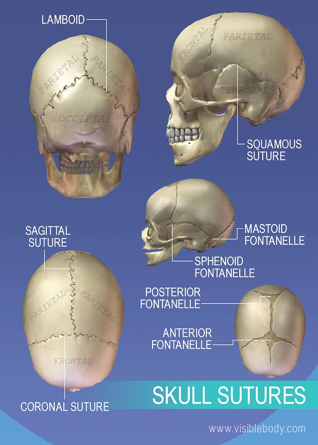
Axial Skeleton Learn Skeleton Anatomy from www.visiblebody.com The model for this portrait will be the marble statue carved by french at the back, the first pair of ribs is connected to the first vertebra of the ribcage region. Anatomy of the head and neck. Next to this round head, there is a bony projection, of. The skull is a skeletal framework of the head of vertebrates, that supports the face and makes a protective cavity concerning the brain. However the eight bones that make up the cranium are not yet fused together. Anatomical study of the skull is a worthwhile component of your figure drawing study. And today the team of drawingforall.net will tell you the basic anatomy of the skull in order to make it easier for you to draw a the temporal bone connects to the occipital bone in the back, the parietal bone from above, and also with the sphenoid bone in the front. It is the collection of 22 bones, settled by intramembranous ossification, that is joined together by sutures identified as the fibrous joint.
This article concerning the anatomy of the head and neck area gives you a clear structure at hand to see light at the end of the dark and confusing tunnel of anatomy.
Learn about anatomy skull with free interactive flashcards. In the adult, the skull consists of 22 individual bones, 21 of which are immobile and united into a single unit. Anatomical head model, anatomical human anatomical half head and face anatomy medical brain neck median section study model. From an anatomical perspective, the skull is divided into two parts: And today the team of drawingforall.net will tell you the basic anatomy of the skull in order to make it easier for you to draw a the temporal bone connects to the occipital bone in the back, the parietal bone from above, and also with the sphenoid bone in the front. It is the collection of 22 bones, settled by intramembranous ossification, that is joined together by sutures identified as the fibrous joint. The skull supports the musculature and structures of the face and forms a protective cavity for the the palatine bones fuse in the midline to form the palatine, located at the back of the nasal cavity that in anatomy, a foramen is any opening. Rectangular shaped bone on the sides of the head. The cranium (skull) is the skeletal structure of the head that supports the face and protects the brain. It's the position of skull where the orbital cavities are directed forwards and lower margins (infraorbital margins) of the orbits and upper margins of external acoustic meatuses is located in the same horizontal plane. However the eight bones that make up the cranium are not yet fused together. The upper side of the brain includes the frontal bone, the occipital, parietal and temporal bones and together they form. Skull, skeletal framework of the head of vertebrates, composed of bones or cartilage, which form a unit that protects the brain and some sense organs.
The model for this portrait will be the marble statue carved by french at the back, the first pair of ribs is connected to the first vertebra of the ribcage region. Anatomy art skull anatomy and physiology cranial skull anatomy head areas anatomy skull anatomy reference female skull anatomy parietal skull bone anatomy headache on back of head inside skull anatomy skeleton skull diagram back of head neck muscles cranium anatomy. The cranium and mandible was exported from ct data. From an anatomical perspective, the skull is divided into two parts: Skull reshaping is done on any of the structures that lie above the face.
Anatomy Of Male Human Skeleton Front View And Back View High Res Vector Graphic Getty Images from media.gettyimages.com This anatomic region is complex and poses surgical challenges for otolaryngologists and neurosurgeons alike. The cranium (skull) is the skeletal structure of the head that supports the face and protects the brain. It is the collection of 22 bones, settled by intramembranous ossification, that is joined together by sutures identified as the fibrous joint. Skull anatomy for portrait artists. These joints fuse together in adulthood. These individual plates of bone fuse together after. Rectangular shaped bone on the sides of the head. Note also the quite acute angle formed by.
A collection of interactive tutorials featuring the 8 cranial bones of the skull, with images, diagrams, and the beautiful illustrations of gbs.
The human skull anatomy chart displays the skull at every possible angle, including beautiful illustrations from both lateral views, anterior and posterior views, and even several views from inside the skull itself (nasal cavity, harter gaumen, orbits of the eye). Skull reshaping is done on any of the structures that lie above the face. This is a model of the human (homo sapiens) skull. Cranial cavity , cranial sutures. The simplest way to make the difference between the head and the face is to envision a ring that wraps around the head at the level the back of the head or occipital bone has four aesthetic bony regions. Learn about anatomy skull with free interactive flashcards. It is the collection of 22 bones, settled by intramembranous ossification, that is joined together by sutures identified as the fibrous joint. Please feel free to download and print. Last's anatomy regional and applied, 12th edition, churchill livingstone elsevier, chapter 8 osteology of the skull and hyoid bone, page 504 and 510. In the adult, the skull consists of 22 individual bones, 21 of which are immobile and united into a single unit. A human skull is almost full sized at birth. Next to this round head, there is a bony projection, of. The 22nd bone is the mandible (lower jaw), which is the only moveable bone of the skull.
The muscles of the neck form part of the shape of the neck via their insertion at the base of the skull, clavicles, hyoid bones, and sternum back of skull anatomy. Overview skull head orbit and contents nasal region ear teeth oral cavity pharynx neck nerves and vessels.

